


Robert J. Schwartz Memorial Lecture
The annual Robert J. Schwartz Memorial Lecture was established by Dr. Gail Ross in memory of her husband, Mr. Robert J. Schwartz, a compassionate and dedicated former member of the Board of Trustees who passed away in 1997. The lecture in his memory seeks to bring experts in the field of reading and reading disabilities to the School each spring.
Past Robert J. Schwartz Memorial Lectures
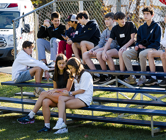
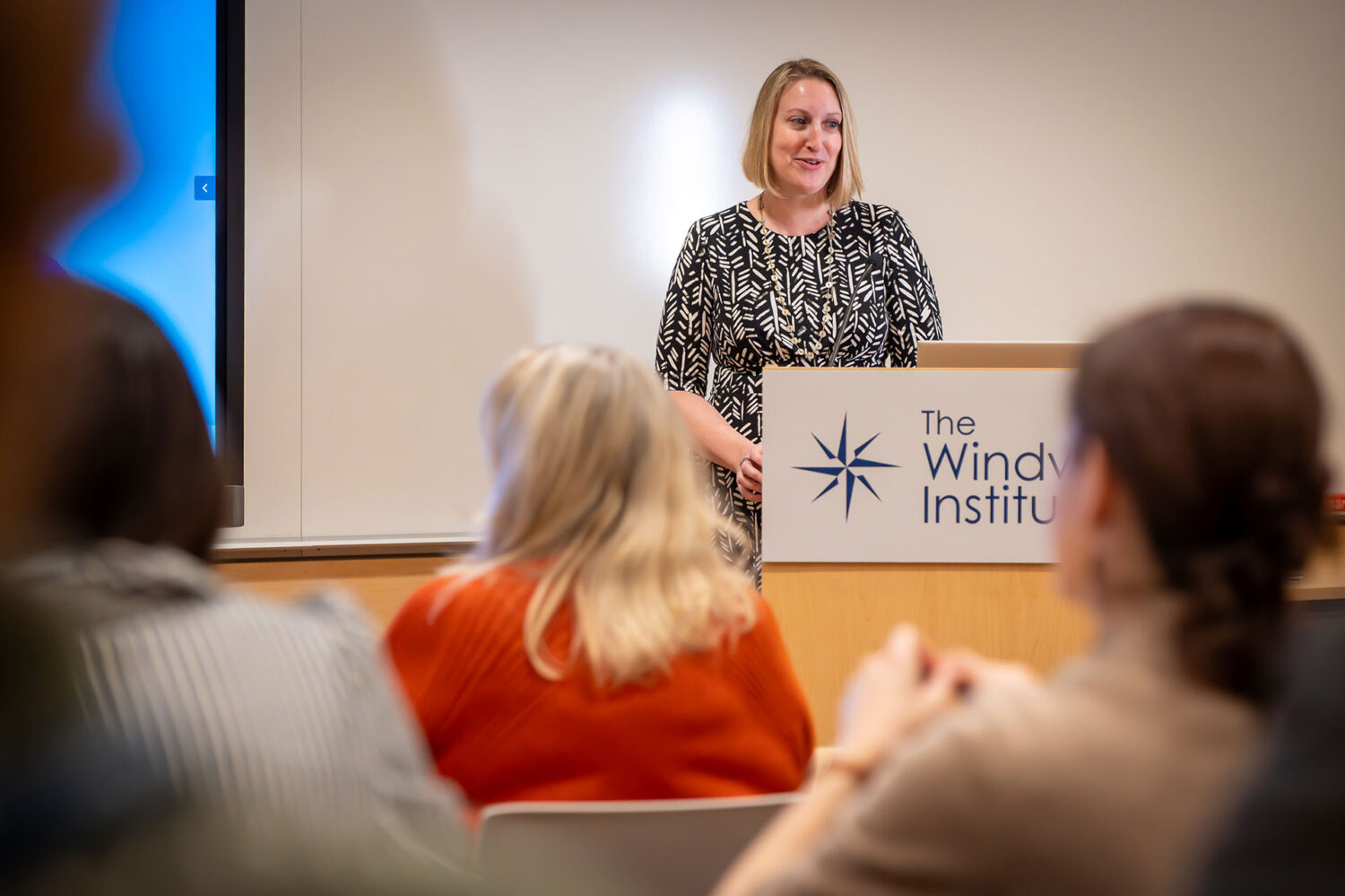
The Windward Institute presents two free educational lectures open to the public in the spring and fall each year
Nadine Gaab, PhD, is a prominent researcher and international speaker on reading development. Her work focuses on developmental cognitive neuroscience, particularly in language-based learning disabilities.
Dr. Gaab focused on learning differences in reading acquisition within a learning disability framework. She presented results from longitudinal behavioral and neuroimaging studies that characterize differences in learning to read as a complex outcome of cumulative risk and protective factors interacting within and across genetic, neurobiological, cognitive, and environmental levels from infancy to adulthood.
The Spring 2020 Robert J. Schwartz Memorial Lecture, "Early Identification of Dyslexia: Research to Practice" with Hugh Catts, PhD, has been rescheduled to Spring 2021.
The Windward School and The Windward Institute were looking forward to welcoming Hugh Catts, PhD, to present his lecture "Early Identification of Dyslexia: Research to Practice."
Due to the incredibly complex and rapidly evolving situation created by COVID-19, the Robert J. Schwartz Lecture that was to be held on April 14 was canceled. Dr. Catts has agreed to present at our 2021 Robert J. Schwartz Lecture.
Educational neuroscience is an emerging field of research that draws upon the disciplines of cognitive neuroscience, education, and psychology, with the goal of examining neurobiological processes as related to education. In this lecture, the neural mechanisms of reading, mathematics, and attention were discussed as well as insights about how this emerging field can influence instructional practice. In addition, neurobiological approaches that may inform and refine our understanding of how to identify and treat reading difficulties were discussed.
On Wednesday, April 26, 2017, nearly 500 people attended the annual Robert J. Schwartz Memorial Lecture presented by internationally recognized neuroscientist Dr. Guinevere Eden. In her presentation on “Decoding the Reading Brain: Lessons from Brain Imaging,” Dr. Eden described how she and her colleagues were the first to apply functional MRI (fMRI) to the study of dyslexia, and they have since used this brain-imaging technology to visually map the functions of the brain. Advances in brain imaging technology have enabled researchers to use non-invasive tools to understand the cognitive processes for language and reading acquisition. While fMRI scans have broadened our understanding of dyslexia, they have also helped neuroscientists discover the positive results of effective intervention to remediate dyslexia.
Dr. Eden’s research has significantly contributed to mapping the reading brain as well as understanding the neural correlates of dyslexia. At the lecture, she shared studies that show the differences in language and reading acquisition across different writing systems and oral languages. She also shared other studies that demonstrate how brain activity changes based on the age of when a person learns to read. For example, as young children learn how to read, the studies show increased activity in the area of the brain related to phonological awareness. These brain images have shown distinct differences in brain mapping based on a person’s native language, writing system, and level of reading development.
In dyslexia research, brain imaging has been integral in supporting and expanding existing behavioral studies. Dr. Eden presented many fMRI studies that charted differences in the reading brains of people with and without dyslexia. According to brain imaging studies of people with dyslexia, certain areas of the brain are under-activated during reading tasks. More recently, other brain imaging studies have shown that people with dyslexia also exhibit under-activated areas of the brain during mathematical procedures tasks.
Neuroscientists have also used brain imaging to demonstrate how effective interventions and instructional practices have changed the brains of struggling and pre-literate readers.In various studies of adults and children with dyslexia, fMRI imaging showed that new areas of the brain were activated after an intensive, structured, and multisensory intervention, demonstratingfurther evidence of neuroplasticity in the brain. Therefore, Dr. Eden emphasized the importance of seeking early intervention for struggling readers. Although brain imaging is not intended for individual diagnosis, aggregated findings from participants across a multitude of studies have provided new insights for neuroscience and education. Future brain imaging research and collaboration between scientific institutions and educational communities will continue to deepen our understanding of dyslexia and the developing reading brain. – Danielle Scorrano, The Windward School Research Associate
Fumiko Hoeft, M.D., Ph.D., is an internationally recognized cognitive neuroscientist. Dr. Hoeft is Associate Professor of Child & Adolescent Psychiatry, Dyslexia Center, and Director of the USDF Hoeft Laboratory for Educational Neuroscience (brainLENS.org). She received research training at Harvard, UCLS, Caltech and Stanford.
In her presentation, Dr. Hoeft discussed how neuroscientists, educators, and practitioners have the goal of helping children reach their full potential and, ultimately. lead fulfilling lives. She discussed her current research program that focuses on brain development and the brain mechanisms that underlie the acquisition of skills such as language and reading. Dr. Hoeft's program also conducts research into other educationally relevant concepts suh as motivation, Mindset Theory, grit, and stereotype threat.
"How Cognitive Neuroscience May Contribute to Helping People with Dyslexia"
Cognitive neuroscience, via neuroimaging, has revealed brain differences associated with difficulty in learning to read, and also brain plasticity associated with effective instruction. But how can basic science knowledge about the brain translate to actual help? Recent advances in brain research may begin to help in practical ways, such as more accurate early identification, diagnosis, and prognosis.
John Gabrieli is the Director of the Athinoula A. Martinos Imaging Center at the McGovern Institute for Brain Research at MIT. He is an Investigator at the Institute, with faculty appointments in the Department of Brain and Cognitive Sciences and the Institute for Medical Engineering & Science, where he holds the Grover Hermann Professorship. He also has appointments in the Harvard Graduate School of Education, and in the Department of Psychiatry at MGH. His area of research is human cognitive neuroscience in which he studies the brain basis of memory, language, and thought. His recent research has focused on brain development in children and adolescents, including studies of dyslexia, ADHD, and autism. He is the first or senior author of over 250 papers published in peer-reviewed scientific journals, including Science and Nature.
Dr. Gordon F. Sherman, the executive director of The Newgrange and Laurel Schools in New Jersey and The Newgrange Education Center, gave an informative presentation to Windward parents, faculty, and educators from the broader community.
His topic for the evening, "Welcome to the Future: Where Diverse Brains Thrive," detailed the diverse ways in which the human brain functions. Understanding the basic architecture of the brain, Dr. Sherman stated, is very important.
Dr. Gaab focused on learning differences in reading acquisition within a learning disability framework. She presented results from longitudinal behavioral and neuroimaging studies that characterize differences in learning to read as a complex outcome of cumulative risk and protective factors interacting within and across genetic, neurobiological, cognitive, and environmental levels from infancy to adulthood.
"How Can Knowledge of the Reading Brain Advance Instruction and Change our View of Dyslexia"
This presentation provided an overview of current work in the neurosciences on the development of the reading brain's new circuit and the importance of addressing the many components within this circuit in our teaching, prediction and diagnosis of dyslexia. A conceptualization of dyslexia that emphasizes the advantages conferred, along with disadvantages will be described.
Maryanne Wolf is the John DiBiaggio Professor of Citizenship and Public Service and Director of the Center for Reading and Language Research in the Eliot-Pearson Department of Child Development at Tufts University. She received her doctorate from Harvard University. Among her awards for teaching and research are Distinguished Professor of the Year award from the Massachusetts Psychological Association, Teaching Excellence Award from the American Psychological Association, Fulbright Fellowship, Distinguished Researcher Award from Tufts University, Norman Geschwind Lecture Award and the Samuel T. Orton Award from the International Dyslexia Association, and the NICHD Shannon Award for Innovative Research, the basis of the RAVE-O reading intervention program.
Wolf is the author of over 140 professional articles and also a book for the general public, Proust and the Squid: The Story and Science of the Reading Brain, which is translated into thirteen languages and an audio version. Dr. Wolf’s research interests include reading interventions, imaging studies of the developing reading brain, the genetic basis of dyslexia, early prediction, fluency and naming speed, cross-linguistic studies of reading, and the future of the reading brain in a digital culture. Her most recent research involves the development of a reading tablet to help children develop literacy both in regions of the world where there are no schools and in schools in the U.S. and other countries where there are too few teachers and resources.
The Windward School and The Windward Institute were looking forward to welcoming Hugh Catts, PhD, to present his lecture "Early Identification of Dyslexia: Research to Practice."
Due to the incredibly complex and rapidly evolving situation created by COVID-19, the Robert J. Schwartz Lecture that was to be held on April 14 was canceled. Dr. Catts has agreed to present at our 2021 Robert J. Schwartz Lecture.
The Windward School was honored to host Ken Pugh, Ph.D., a distinguished research scientist in the field of language, reading and reading disabilities, at this year’s Robert J. Schwartz Memorial Lecture. Held on the evening of Wednesday, April 6 at the Red Oak Lane campus, Dr. Pugh delivered an animated and detailed presentation to an audience of 450 people, including members of the Windward faculty and administration, as well as parents, physicians and educators from area schools.
Dr. Pugh serves as President, Director of Research and Senior Scientist at Haskins Laboratories, an institution dedicated to the study of the biological basis of language. The laboratories are affiliated with both the University of Connecticut and Yale. Dr. Pugh also works as the director of the Yale Reading Center, which studies the neurobiological basis of reading disability and development. His lecture, Neuroimaging Studies of Reading and Language Development: An Update on Recent Findings, explained the manner in which scientists study the brain and the ways in which it is activated during the reading process by both typical and dyslexic readers.
In addition to Dr. Pugh, two of his assistants from Haskins Laboratories, Beth Eaton and Annie Stutzman, paid a visit to the School. Ms. Eaton and Ms. Stutzman spent the day at the School meeting with members of the administration to discuss Windward’s research-based program. The pair also observed reading and writing classes at both the Lower and Middle School campuses. Both women had strong praise for the School’s curriculum and faculty.
Dr. Pugh himself spent the evening of his lecture discussing the intricacies of using fMRI (functional magnetic resonance imaging) to study brain activity. He detailed the manner in which the dyslexic brain differs from the typical brain, and how research-based, multisensory intervention of the type given at Windward changes brain activation patterns. Dr. Pugh explained how fMRI’s are producing positive changes in the ways in which researchers understand and ultimately treat dyslexia and language-based learning disabilities. A dedicated scientist with a flair for presenting complex topics in an engaging and applicable manner, Dr. Pugh spoke in great detail about the advancements being made in the field of reading disability research. The School was quite fortunate to host such a distinguished scholar to campus.